


-
自16世纪末第一台光学显微镜在荷兰问世以来,光学显微技术经历了不断的革新。它已经从简单的单透镜成像装置发展成为了一种极为重要且精密的观察与计量科学仪器,广泛地应用于生物、化学、物理、冶金、酿造、医学等各种科研活动,对人类的发展做出了巨大而卓越的贡献。特别是在生命科学领域,光学显微技术引领着人类打开微生物世界的大门,为生命科学研究与现代临床疾病诊断提供有力的影像学依据,成为推动生命科学和基础医学进步不可或缺的重要工具。
自诞生来,光学显微镜的发展一直所面临着一个巨大挑战—“分辨率”。自恩斯特·阿贝(Ernst Abbe)1873年发表著名的阿贝分辨率极限公式以来[1],人们一直认为显微镜的分辨率只有光波长的一半。对可见光而言,这个数值最小约为200 nm。于是,物理学家很自然地将思路转向波长更短的射线,发明了电子显微镜和X射线显微镜。这两种显微镜都能达到纳米甚至更高的分辨率。可惜这两种显微镜都无法对生物活体进行有效且无损的观测。1962年,下村修(Osamu Shimomura)[2]从生活在美国西海岸近海的翼钟水母身上提取、鉴定出了绿色荧光蛋白。这种绿色荧光蛋白使通过设备肉眼观察、追踪被标记的活体细胞成为可能。荧光显微镜[3]、激光共聚焦显微镜[4]、全内反射荧光显微镜[5]、双/多光子荧光显微镜[6-7]、光片显微镜[8-9]等的问世极大地促进了生命科学研究水平的进步。这些技术允许检测非常微弱的荧光信号,并以高特异性揭示样品的结构和功能特性,为生物与医学研究带来了一场革命。2008年,诺贝尔化学奖授予了绿色荧光蛋白,标志着以荧光蛋白为基础的现代分子成像技术已经成为了当代生命科学研究中与显微镜的发明相提并论的最重要的工具之一。更加让人意想不到的是,荧光分子开关效应的发现和单分子探测技术的出现,使连续观测单个荧光分子行为而非其集合平均成为可能。受激发射损耗显微镜(STED)[10]、光敏定位显微镜(PALM)[11]、随机光学重建显微镜(STORM)[12]等超分辨成像技术利用荧光分子的受激辐射原理与光开关效应,绕开了一百多年来似乎一直被认为是无法突破的阿贝衍射极限,将光学显微技术带入到纳米尺度。2014年,诺贝尔化学奖授予了超分辨率荧光显微技术,再一次展现了光学显微技术在人类发展历程中以及未来发展方向上的重要地位。
截止目前,荧光显微技术已逐步实现从2D宽场到3D共聚焦/双光子与超分辨的跨越。然而这类方法仍需要挑选具备合适发色团的样本,或者对样本进行荧光标记处理以提高成像的特异性和衬度。实际上,无论是哪种标记方法,都存在一定的局限性[13]:首先,荧光染色本身就是个复杂耗时的过程,且荧光标记物不可避免地对细胞机体的正常代谢活动产生不利影响;其次,激发光对细胞的光损伤较大,荧光基团还存在光漂白的问题,这些阻碍了对细胞的长时间连续观测;最后,细胞内某些重要组分,如小分子和脂类的特异性不高,荧光标记往往只能突出部分生物分子而难以获得细胞整体全貌。
在研究活细胞的动态过程及其各项生理活动时,无标记(Label-free)显微是一种最为理想的探测手段[14-15]。当光通过几乎透明的物体(如细胞)时,其振幅几乎不变;然而透射光的相位则包含了关于样品的重要信息,如形貌与折射率分布。1932年,Zernike基于这种想法发明了相差显微镜:利用光的衍射和干涉特性并根据空间滤波的原理改变物光波的频谱相位,成功将相位差转换成振幅差,从而极大地提高了透明相位物体在光学显微镜下的可分辨性[16]。相衬法的发明具有划时代的意义,Zernike因此获得1953年的诺贝尔物理学奖。同期,各类无标记显微技术也得到了快速发展,例如用来观测散射物体的暗场显微镜,用来观测晶体的偏振显微镜等,这些技术已在各类无标记成像研究应用中无处不在。然而,这些技术从发展初期到现在并没有经历太大的革新,且成像分辨率仍然受到阿贝衍射极限的限制,无法实现超分辨成像。不论是理论与技术方面的发展与荧光显微成像相比都显著滞后。如何在“无标记”的前提下实现超分辨显微成像,是目前显微镜技术发展亟待突破的一大重点与难点。
1972年,近场扫描光学显微技术(Near-field scanning optical microscopy, NSOM)诞生了,其基本原理是利用孔径小于入射光波长的针孔对样品表面倏逝波进行收集。倏逝波是一种沿介质界面传播、振幅垂直于界面的电磁波,其在传播过程中可携带物体的高频率亚波长空间信息[17]。NSOM技术使得人们首次能够突破光学衍射的限制,进行亚波长超分辨成像。虽然NSOM技术的成像分辨率远优于传统光学显微镜,但倏逝波的振幅在与样品表面垂直的传播方向上会随距离的增大呈指数衰减,在该方向上的传播距离只有入射波长量级。因此,必须在待测样品的近场区探测,通过探针逐点扫描的方法进行成像,这也使得整个系统复杂且成像缓慢,无法实现实时的生物检测,极大限制了该技术的应用[18]。
-
2009年,韩国浦项科技大学(Pohang University of Science and Technology)的J.-Y. Lee等人首先报道了微米透镜具有超分辨成像能力[19]。该团队通过化学方法制备了直径0.05~3 µm、厚度小于0.8 µm的C28H24O8 (CHQ)平凸透镜(n=1.50),并利用CHQ微透镜成功在高倍物镜(100×, 0.9 NA)下对间隔250 nm和220 nm的Pd/Cr金属线条进行了显微成像,从而证明了CHQ微透镜的分辨能力已超越了Abbe分辨率极限,可达到0.34λ/NA。他们用时域有限差分法(Finite difference time domain, FDTD)对CHQ微透镜的近场聚焦现象进行了仿真研究,发现用FDTD法算出的微透镜焦距远小于用光线追迹法算出的焦距(图1(a))。2011年,英国曼彻斯特大学(University of Manchester)的Z. Wang等人[20]利用直径2~9 µm的SiO2微球(n=1.46)结合80×,0.9 NA的物镜成功在卤素灯照明下实现了50 nm微结构的观测,其成像系统分辨率能达到λ/8~λ/14,放大率介于4~8倍之间,并通过该成像系统成功分辨出了线宽200 nm、间距100 nm的光栅结构,以及直径~50 nm、间距~50 nm的非周期圆孔结构(图1(b))。Wang等人通过将直径4.87 µm的SiO2微球与直径2.5 mm、高度0.5 mm的SiO2固体浸没透镜进行对比发现微透镜的分辨能力远超固体浸没透镜。同年,浙江大学刘旭团队发现当在微球上滴加酒精后,酒精液膜厚度会因为蒸发现象而逐渐减小到与微球直径相当或者小于微球直径,此时微球的超能分辨成像质量会得到显著提升。利用该方法,他们通过直径3 µm的SiO2微球对蓝光光碟表面条纹进行了对比度增强超分辨成像[21]。2012年,美国北卡罗来纳大学夏洛特分校(University of North Carolina at Charlotte)的A. Darafsheh发现直径为2~220 µm的BaTiO3微球(n~1.9~2.1)在液体完全浸没时同样具有超分辨成像能力,其分辨率最高能达到λ/7,当微球的直径大于50 µm时,其分辨率约为λ/4。他们利用异丙醇完全浸没的BaTiO3微球成功实现了对间距150 nm、直径120 nm、高度30 nm的周期性金点阵的超分辨成像[22]。此后,聚苯乙烯[23]、TiO2-BaO-ZnO微球[24]等不同材料微球均被证实具有超分辨成像能力,并相继衍生出了微球辅助共聚焦显微成像技术[25]、扫描微球超分辨成像技术[26-27]等相关技术。
近年来,国内众多课题组也针对此方向开展了相关研究工作:中国科学院沈阳自动化所刘连庆团队将微球与原子力显微镜悬臂粘结,结合图像拼接技术实现了非接触大面积扫描超分辨成像(图1(c))[26],并将该方法拓展到微操控领域提高了可视化微操控的精度[28]。其团队还研究了级联型微透镜组的成像特性,并实现了无浸没高质量超分辨成像[29]。复旦大学武利民团队通过自组装技术将粒径为15 nm的TiO2颗粒组装成直径10 µm的透镜,并实现了对45 nm微结构的光学显微成像(图1(d))[30]。暨南大学李宝军团队利用光力操控C10H7Br液滴,通过改变液滴外形可对其超分辨成像特性进行实时调控(图1(e))[31]。南京师范大学叶永红课题组研究了SiO2微球和BaTiO3微球的远场成像特性,建立了成像视场、放大率和物距之间的关系[32–34],成像系统景深和光子射流长度之间的关系[35],并采用半浸没结构提高了微球透镜的数值孔径和分辨能力[36],改善了成像畸变[37],通过在基板表面制备高反膜提高了成像对比度[38]。2020年,叶永红课题组发现在样品表面沉积金属-介质膜也可提高微球成像系统的分辨率和对比度[39](图1(f)),并对微球超分辨成像的物理机制进行了研究[40]。

Figure 1. (a) CHQ microlens-assisted super-resolution microscopy[19]; (b) SiO2 microsphere-assisted super-resolution microscopy[20]; (c) Scanning microsphere super-resolution microscopy[26]; (d) Self-assembled TiO2 particles for super-resolution microscopy[30]; (e) Liquid droplet-assisted super-resolution microscopy[31]; (f) Surface plasmon resonance-enhanced super-resolution microscopy[39]
根据上述工作可看出,相比于NSOM技术,微球超分辨显微成像技术仅需在原有的显微光学系统中加入直径几微米至几百微米的透明介质微球就能够成倍提高传统光学显微镜的成像能力。此外,它还具有宽场成像和无标记成像等显著优点,能在宽谱照明条件下工作,并且可与市场上成熟的显微镜产品相兼容,因此极具研究价值与应用前景,发展潜力巨大。
此外,微球超分辨技术在光学测量领域也具有潜在的应用价值。2016年,北京工业大学王大勇团队[41]将微球透镜用在数字全息显微技术中,对一维和二维光栅结构进行了测量,并发现加入微球可显著提升数字全息系统的横向分辨率。该项工作将微球的应用从传统二维成像领域拓展到了三维测量领域。2018年,法国斯特拉斯堡大学(University of Strasbourg)的A. Leong-Hoi等人[42]将直径24 µm的钠钙硅微球(n=1.50)引入相移干涉显微系统中,并利用该系统对多种样品的三维轮廓进行了测量,将干涉显微系统的横向分辨率提升了4倍,并成功对周期为300~400 nm的硅基光栅结构、粒径为200~300 nm的银纳米颗粒、周期为900 nm的波浪状微结构进行了三维轮廓测量。2019年,中国科学院光电技术研究所胡松课题组[43]将正弦调制的结构光通过直径20 µm的SiO2微球聚焦到观测样品表面,以提升显微测量系统的分辨率。通过该方法,他们成功对高度150 nm、线宽220 nm、周期500 nm的硅基光栅结构样品进行了超分辨三维显微轮廓测量。
与微球超分辨成像的实验现象发现与应用技术开发快速发展形成鲜明对比的是,微球超分辨成像的物理机制现阶段仍存在一些争议,尚未有最终定论[44]。Wang认为微球能够超分辨的原因在于其具有光子纳米射流现象。既然微球能将入射光波进行超聚焦,会聚到亚波长尺度,那么根据光路可逆原则,超分辨精细结构自然也应该能被微球传播到远场,而其成像过程则与传统透镜一样,都是通过光的折射,将微球下方样品的光信号传输到远场,并最终被高倍物镜接收[20]。Hao和Yang认为携带物体高频信息的倏逝波(Evanescent wave)本身并不能传播到远场,但微球可与其在近场区域内发生耦合,正是该近场区域内的耦合作用才是微球具有超分辨成像能力的关键,而通过调节匹配介质层的形状和对样品表面进行金属化等处理,可使得耦合作用得到增强,从而提高微球成像质量[25,39]。Duan和Hoang通过仿真研究发现回音壁共振模式(Whispering gallery mode,WGM)可提高微球的分辨率,因此他们认为微球的超分辨能力源于WGM共振现象[45-46]。
虽然上述工作给出了许多具有启发性的研究成果,但是目前尚没有一套公认的理论能够完整且准确解释微球透镜的超分辨效应。这致使该领域目前仍缺少有效的方法来对微球透镜成像性能进行定量优化以进一步提升其成像潜能。在此背景下,笔者们认为有必要对现有的与微球透镜近场聚焦及远场成像机理与模型相关的研究成果进行梳理与归纳,分析现有工作的意义与不足,指出该领域未来需要着重解决的重点问题,并对微球成像技术未来的发展方向给予展望。
-
微球透镜近场聚焦特性方面的研究目前主要是通过求解麦克斯韦方程组计算出微球附近的电磁场物理参数和光强空间分布,由于光波的衍射现象在微透镜附近会变得非常显著,因此如标量衍射理论、光线追迹等经典仿真手段不再适用。
在近场域的成像仿真需要根据矢量衍射理论才能准确获得光场的物理参数,目前微球成像研究中常用的商业软件有基于时域有限差分法的Lumerical FDTD Solutions,基于有限元法的COMSOL Multiphysics以及基于有限积分法的CST MICROWAVE STUDIO,等等。因为近场域的全波矢量仿真对于电脑内存要求很高,所以大部分课题组都采用二维仿真的手段来进行计算,三维仿真通常只能在一个微小区域内进行。远场域的成像仿真则可通过标量衍射理论里的角谱传播等方法进行快速计算。因此,可以看出,近场和远场域的光学仿真手段的主要区别在于是否需要求解麦克斯韦方程组,并由此导致后期算法的不同以及相应计算量的差异。
目前大部分课题组都是将微球近似为一个圆柱体,研究该圆柱体一个剖面上的光场聚焦特性,并以此近似看成为微球的聚焦特性。入射光波经微球会聚后所形成的高强度光场又可根据其空间结构分为两大类:具有轴对称结构的光子纳米射流(图2(a))、具有弯曲结构的光子钩(图2(b))。

Figure 2. (a) A photonic nanojet and (b) a photonic hook; (c) Super-resolution imaging technology based on photonic hooks[47]
-
研究表明,当电磁波穿过具有与其波长可比拟尺寸介质微粒时会产生一种特殊的电磁波会聚现象,并在微粒背面阴影处产生狭长的光场,由于该光场的光强空间分布形似飞机引擎喷射出的气流,因此又被称为光子纳米射流(Photonic nanojet, PNJ)。光子纳米射流具有以下几个重要性质[48]:(1)光束横截面的半高全宽(Full width at half maximum, FWHM)可以小于光学衍射极限,并沿传播方向传播2λ以上;(2)当微粒与环境的折射率比值小于2时,直径在2λ~40λ范围内的介质微柱或微球都可激发光子纳米射流;(3)光子纳米射流的强度远高于激发光的强度;(4)位于光子纳米射流中的纳米颗粒的背向散射信号可被增强几个数量级。正因为光子纳米射流具有上述优点,其在微纳制造、粒子探测及操控以及超分辨显微成像等领域具有良好的应用前景。
PNJ的亚波长聚焦现象能够显著提高光学成像系统的分辨能力,实现无标记超分辨显微成像功能,而微球透镜的成像特性也与其PNJ的特征参数有着密切关联[44]。2016年,瑞士洛桑联邦理工大学(École Polytechnique Fédérale de Lausanne)的H. Yang等人发现了微球PNJ的FWHM越小其分辨率越高[49]。他们利用直径4.2~11.8 µm的BaTiO3微球(n=1.92)对线宽120 nm,间隔100 nm的光栅结构在水镜(40×,0.8 NA)观测下成功进行了超分辨成像,并发现直径6~7 µm的BaTiO3微球所成的像具有最高的放大率(M~5.95×),通过算出了微球成像系统的点扩散函数(Point spread function, PSF),发现直径~6 µm的BaTiO3微球分辨率最高,而仿真结果也证实了该尺寸微球的PNJ具有最小的FWHM。2017年,南京师范大学叶永红团队发现微球成像系统的可成像范围随PNJ长度的增加而变长[36]。他们通过将微球包裹在聚二甲基硅氧烷(Polydimethylsiloxane, PDMS)透明薄膜里,在落射式明场显微成像系统下,对线宽为200 nm、间距为100 nm的光栅结构和周期为960 nm的六角密排(Hexagonally-close-packed, HCP)结构进行成像,发现当微球直径从5 µm增大到50 µm时,其可成像范围从4 µm增加到25 µm,利用FDTD仿真发现微球的PNJ长度也随其直径的增加从2.9 µm逐渐增大到7.1 µm。2017年,美国宾夕法尼亚大学(University of Pennsylvania)的A. Darafsheh发现微球PNJ的FWHM越小,其成像质量越高,而当微球与环境之间折射率比值固定时,通过FDTD仿真可发现PNJ的FWHM随微球折射率的增大而减小,因此BaTiO3微球(n~1.90)比SiO2微球(n~1.45)具有更优异的成像性能[50]。2021年,暨南大学李宝军团队发现生物细胞同样可以产生PNJ并实现超分辨成像的效果,他们利用光镊技术操控直径0.5~40 µm的人体脂肪细胞(n~1.52),成功对细胞间和细胞内部的细胞骨架、溶酶体和腺病毒荧光信号进行了超分辨探测和成像[51]。
-
2018年,英国班戈大学(Bangor University)王增波课题组和俄罗斯托木斯克理工大学(National Research Tomsk State University)Minin课题组通过理论计算发现了一种自弯曲光子射流[52],2019年又通过实验进行了验证[53]。该新型光子射流最窄处半宽约为0.45λ,最强处光强可接近入射光强的15倍,最大弯曲角度可达35°。如图2(a)所示,由于该光子射流的光场空间分布形似弯钩,因此又被称为光子钩 (Photonic hook)。目前自弯曲光子射流的激发方式主要有四种:(1)使光波与非对称粒子相作用,例如基于梯形粒子的透射激发模式[52-53]和基于台阶镜面的反射激发模式[54];(2)使用非对称入射光场,例如通过遮光板使介质微柱部分区域处于挡板阴影处,在透射模式下可激发自弯曲光子射流[55-56];(3)使光波穿过变折射率粒子,例如Minin课题组利用Janus立方体激发自弯曲光子射流[57],南方科技大学邵理阳和深圳大学宋军课题组利用Janus圆柱体激发自弯曲光子射流[58];(4)使光波与特殊形状对称粒子相作用,例如基于孪生双微柱结构的透射激发模式[59],该模式可同时产生两个自弯曲光子射流。
2021年,南京师范大学汤芬等人利用FDTD法对补丁粒子的近场聚焦特性进行了仿真研究[60]。在该项工作中,折射率为1.90的圆柱形BaTiO3微粒被分别放置在空气(n=1.00),水(n=1.33)和香柏油(n=1.52)中,微粒表面覆盖有100 nm厚的银膜。由于该银膜就像在微粒表面打上补丁一样,因此这类微粒统称为补丁微粒(Patchy particle)。当用波长为550 nm的单色平面波对补丁BaTiO3 微粒进行照射时,会在其背面阴影侧形成一道弯曲光束,即光子钩。微粒的光子钩与其粒径大小和表面银膜覆盖率有着密切关系。当其表面1/2面积被银膜覆盖时,光子钩弯曲角度会随粒径的尺寸的减小而增大,因此直径35 µm和10 µm的微粒产生的光子钩所具有的弯曲角度分别为15.4°和20.2°。而当银膜覆盖率减少到1/4时,直径1 µm的微粒仍然能将平面波会聚成为光子钩。利用该方法,他们发现补丁BaTiO3微粒所能产生的光子钩最大弯曲角度~28.4°,光强最强处的半高全宽~0.36λ。
2021年,南京师范大学尚晴晴等人利用直径~35 µm的补丁BaTiO3微球作为微透镜,对亚波长尺度线状和点状微结构进行了显微成像,并发现补丁微球可显著提高超分辨成像质量[47]。在该实验中,他们首先利用倾斜镀膜法在微球表面蒸镀100 nm厚的银膜,然后通过水流将微球转移到观测样品表面,最后再用倍率20×、NA=0.4的物镜在传统明场落射式显微镜(Axio AX10, Carl Zeiss)下对样品进行观测,成功观测到BaTiO3微球对样品表面微结构所形成的放大的像,达到了超分辨成像效果。通过该补丁微球辅助成像系统,他们对蓝光光碟表面线宽200 nm,间距100 nm的周期性条纹和具有HCP结构的直径230 nm的SiO2颗粒阵列进行了成像,并发现银膜覆盖可使得微球的成像对比度提高约~5倍(图2(c))。此外,他们还利用FDTD法仿真研究了银膜位置对微球近场聚焦特性的影响,发现当覆盖微球表面1/4的银膜与微球顶部夹角为0°~30°之间时,补丁微球的内部会形成弯曲的聚焦光场,即光子钩 (图2(b))。而他们认为光子钩的存在会产生近场倾斜照明的效果,因而可以提高成像质量。
-
虽然通过分析PNJ可以解释微球成像实验中所观测到的部分现象,例如像面位置和放大率的变化。但是一个能将照明光源,样品信息,微球参数,以及后端光学成像系统融合在一起的完善的微球成像理论模型依然缺失。目前研究微球成像理论模型的文献较少,由于计算量的限制,大部分工作都是通过求解麦克斯韦方程组进行二维仿真,模拟微球对相邻点光源的成像,以此来讨论微球成像系统的分辨率[45-46,61–63],将近场全波仿真和远场标量衍射理论相耦合的混合模式的三维仿真技术近期才被提出[64]。
-
2013年,新加坡国立大学(National University of Singapore)和新加坡-麻省理工大学技术联盟(Singapore-MIT Alliance for Research and Technology)的Y.Duan等人曾尝试求解微球对两个非相干点光源成像的米氏解,以此来解释其超分辨成像机理[45]。该项工作所研究的成像系统如图3(a)所示,其中微球直径为4.74 µm,折射率为1.46。Duan等人发现当微球产生光学WGM模式共振时,其分辨能力会得到提升。如图3(b)所示,在WGM共振模式下微球可分辨间距150 nm的两个非相干点光源,当点光源间距减小到100 nm时则无法被分辨。由于该结果与实验观测数据不符,因此Duan等人认为微球的超分辨能力可能源于多种物理机制的共同作用。2015年,新加坡-麻省理工大学技术联盟的T. X. Hoang等人尝试利用平面波展开的多极子算法对直径4.74 µm,折射率1.46的微球成像系统进行理论研究[46],建立包含聚焦和成像两方面的理论模型。Hoang等人同样发现微球在共振模式下具有较高的分辨能力,其分辨率最高可达到0.24λ,理论上可分辨间距为98 nm,发射波长为402.292 nm的两个相邻点光源。

Figure 3. (a) Schematic drawing of the microsphere imaging system used by Duan et al.; (b) The corresponding imaging simulation results[45]
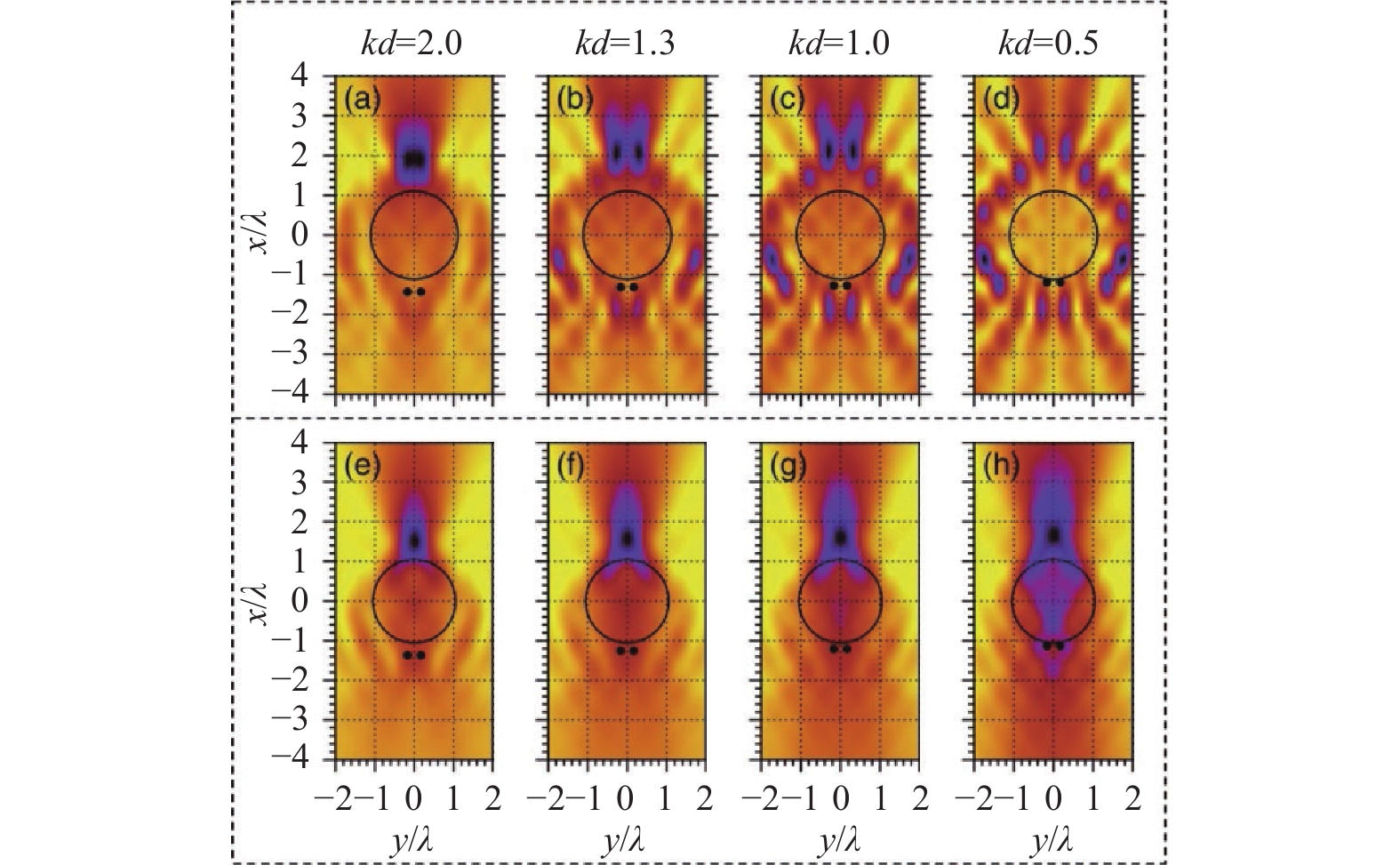
2016年,俄罗斯下诺夫哥罗德国立大学(University of Nizhny Novgorod)的A. V. Maslov和美国北卡罗来纳大学夏洛特分校(University of North Carolina at Charlotte)的V. N. Astratov对半径3.2λ的微球(n=1.4)成像系统进行了二维仿真[61],其观测样品为微球下方0.16λ处的点光源,该方法可以得出与微球分辨率,放大率以及像面位置有关的信息(图4(a)~(e))。Maslov等人发现光源相干性对微球分辨能力有显著影响,微球可以分辨出间距小于λ/16的两个相位相差π的相干点光源,但无法分辨同相位的相干点光源,对于非相干点光源微球的分辨率约为λ/2。2017年,在上述模型的基础上,Maslov和Astratov又研究了米氏共振(Mie resonance)对微球超分辨成像的影响[62],如图5(a)~(h)所示,他们发现在非共振状态下的微球无法分辨出相距λ/3的两个点光源,而在米氏共振状态下,微球分辨能力会得到显著提升,直径在1~2λ之间的微球(n~2)的极限分辨率约为λ/4。

Figure 4. (a) Schematic drawing of the microsphere imaging system; (b) 2D simulation model based on microsphere imaging system; (c) The images of off-axis single point sources formed by microspheres; (d) The images formed by the microspheres for the two point sources distributed symmetrically along the optical axis; (e) The influence of the mode of the point sources (out of phase, in phase, incoherent) on the resolution of the microsphere imaging system[61]

Figure 5. (a)-(d) The images of the two neighboring point sources formed by microspheres when the microspheres are on Mie resonance; (e)-(h) The images of the two neighboring point sources formed by the microspheres when the microspheres are not on Mie resonance[62]
2019年,A. V. Maslov和V. N. Astratov利用该模型对微球聚焦(图6(a))和成像特性(图6(b))进行了比较[63]。他们认为入射光波经微球会聚后所产生的聚焦光场即PNJ,而微球成像系统的PSF即微球对点光源所成的像。仿真结果表明直径约为4 µm的微球(n=1.40)所产生的PNJ的FWHM为~0.32λ,PSF的FWHM为~0.725λ。当微球直径增大到约10 µm时,相应PNJ和PSF的FWHM也逐渐增大到0.6λ和1.15λ,因此PNJ与PSF之间并没有定量关联。

Figure 6. (a) The photonic nanojet of a microlens can be obtained by simulating its focusing performance for plane waves; (b) The point spread function of a microlens can be obtained by simulating its imaging properties for point sources[63]
-
上述方法都属于二维仿真技术,通过将微球简化为圆柱来降低计算量,可是这不可避免的影响了结果的准确性。2020年,台湾大学L. Y. Yu等人首先提出了一种适用于微球成像系统的三维仿真方法[64]。如图7(a)所示,该仿真方法可分为三个部分:首先通过FDTD法在微球附近区域进行全波矢量仿真,再利用算法将微球附近的光场参数耦合到远场,然后在远场区域通过角谱法和反向追迹算法得到样品经光学显微系统后所成的像。通过该方法,Yu等人仿真了放置于空气中的直径4.6 µm,折射率1.46的SiO2微球对间距200~300 nm、波长405 nm的非相干点光源的成像,计算出微球成像系统的分辨率约为250 nm(图7(b))。当利用微球观测金属光栅时,其成像系统的分辨率可达到75 nm,成像畸变也可同时被仿真出来(图7(c), (d))。

Figure 7. (a) The steps for the three-dimensional imaging simulation: (i) The near-field full-wave simulation, the near-to-far-field transformation, the far-field imaging simulation and (ii) the simulation for the formation of virtual images; (b) The simulated microsphere’s imaging performance for the two point sources with various gaps between them; (c) The simulation of microsphere’s imaging for metallic grating structures and (d) the corresponding simulation results show that the metallic lines with 75 nm separation can be resolved by the microsphere; (e) Microsphere imaging system has a higher resolution for metallic samples: (i), (iii) Metallic microstructures; (ii), (iv) Non-metallic microstructures with the same dimension[64]
此外,该仿真技术与其他方法显著不同的地方是可以仿真出样品材料属性对成像效果的影响。如图7(e)所示,当微球对尺寸相同,材料不同的微结构进行成像时,金属样品具有更好的成像效果(图7 (e)中(i),(iii)),而非金属样品的细节则无法被微球分辨(图7 (e)中(ii),(iv))。
-
对于传统显微镜成像系统而言,一般用傅里叶变换和线性滤波来描述图像的生成。对于一个相干成像来说,其为一个复振幅的线性系统;对非相干成像来说,其为一个光强的线性系统。目前大部分对微球成像模型的研究工作都集中在相干与非相干两个极端情形,并侧重于点扩散函数分析,即认为由于成像系统所成图像是系统点扩散函数与物体强度或者复振幅分布函数的卷积,以此对微球透镜的分辨率进行估计。而在一个实际的显微镜成像系统中,通常所面对的是介于相干与非相干的部分相干照明。通过调整显微镜聚光镜的孔径光阑尺寸可以方便地调节照明的数值孔径,即改变照明的空间相干性。显微镜在部分相干照明下的成像特性需借助于Hopkins的光学传递函数理论[65],即四维传递交叉系数来描述。如何将此理论引入微球显微成像,建立其完整的光学传递函数模型,并以此定量分析照明相干性对成像分辨率与对比度的影响,仍是一个值得探究的问题。
-
现有的成像系统模型与仿真方案计算得到的微球透镜分辨率和实验测量的结果有时候能够基本符合。但是由于受到微球种类、成像系统、成像样品、微球直径等方面的影响,实验中获得的成像分辨率和放大倍率都有所不同。此外除了倏逝波以及光子纳米射流效应之外,其他的因素,如金属样品的表面等离激元,微球表面的非理想粗糙结构、成像系统和微球引入的像差与畸变等也会影响微球透镜超分辨成像的实际表现。而完整的微球超分辨成像理论需要考虑微球对入射光的聚焦、样品与聚焦光场的相互作用以及散射光场所成的放大虚像[66]。这说明不能仅仅把微球成像看作传统成像在近场域的简单拓展,倘若能把这些因素全都考虑并进行数学建模,获得完整的微球透镜超分辨成像机制,将有望进一步促进该项技术的工程应用。
-
虽然现有的微球成像方案已取得了良好的超分辨效果,但是引入微球会给后端成像系统带来额外球差,造成纵向和横向分辨率的不同。虽然可以通过图像处理技术进行像差校正,但是当用人眼直接观测时就只能通过改进光学成像系统来减小像差。而微球透镜近场域的作用关系未知,二维和三维点扩散函数和传递函数还未进行深入探究,使得其在光学成像系统设计以及像差校理论上尚不成熟。假以时日,一旦这些问题得以解决,微球超分辨成像技术简单易实现的本征优势,将在微纳米元件的检测材料科学以及生物组织成像等领域迎来广阔的发展空间。
-
微球透镜与光学显微技术相结合,可以突破衍射极限,将显微镜分辨率提升至百纳米甚至更高,该技术发展至今已取得了众多令人瞩目的研究成果。其技术简单,操作方便,易于实现,无标记,分辨率高,具有广阔的应用和发展前景。如今,微球超分辨显微成像技术也逐渐从实验室走向了市场,代表性产品有新加坡国立大学洪明辉院士团队研制的OptoNano-200(Phaos Technology)和英国曼彻斯特大学李琳院士团队推出的NANORO-M(LIG Nanowise)。该领域现阶段的科学研究仍主要集中在微球超分辨成像规律、图像质量的提高、微球的操控方法上,而人们对微球超分辨成像机理经久不息的讨论,以及对微球成像仿真技术的不断尝试,都表明了该领域目前并没有形成一个完善且统一的认知与可靠一致的解释。无论是几何光学、倏逝波、光子纳米射流效应、光学回音壁模式共振以及表面等离激元等等,都无法完全解释实验中出现的一些现象,理论数据和实验结果存在一定出入,完整的成像机制与模型仍在探索中。另外研究者们进行实验时,微球的直径和类别、成像物镜、观测样品、光照条件等实验条件不同,得到的图像放大倍率和分辨率等参数也有所不同,因此评估超分辨图像质量没有统一标准和方法。微球与样品在近场区域是如何相互作用的,微球回音壁模式以及金属表面等离激元是如何对微球超分辨成像性能产生影响的,这些有待回答的问题表明目前仍然还有许多知识层面的空白需要去填补。
微球超分辨显微成像技术未来路在何方?在笔者们看来,“计算光学显微成像”有望成为其未来的重要发展方向之一。不同于传统光学显微成像的“所见即所得”,计算光学显微成像通过对照明与成像系统人为引入可控的编码或者“扭曲”(如结构照明、孔径编码、传递函数调制、探测器可控位移)等并作为先验知识,目的是将物体或者场景更多的本质信息调制到传感器所能拍摄到的原始图像信号中。在解调阶段,基于几何光学、波动光学等理论基础上通过对场景目标经光学系统成像再到探测器这一完整图像生成过程建立精确的正向数学模型,再经求解该正向成像模型的“逆问题”,以计算重构(如相干解调、相位复原、光场调控、压缩感知、单像素重建、反卷积、最优化重建)的方式来获得场景目标的高质量的图像或者所感兴趣的其他物理信息。计算成像将光学调控与信息处理有机结合,有望为开发基于微球超分辨成像技术的新应用提供新思路。但只有明确了微球透镜的超分辨成像机制,理解了微球的作用方式,才能够对微球显微成像系统进行精确的正向建模,实现关键物理机理的数学模型化。这是将微球超分辨显微成像技术从“所见即所得”迈向“计算成像”的关键一步。一旦在此方向上取得了突破,可使其面临的关键问题得以迎刃而解、并快速实现多模态成像并耦合进入多种类型的光学系统,如数字全息显微镜[67]、非干涉定量相位显微镜[68-69]、结构光超分辨荧光显微镜[70]等。微球透镜超分辨显微成像的简单方便结合计算光学显微成像的灵活可控,将有望将微球透镜超分辨显微成像技术的应用空间极大的拓展,在工业检测、材料表征、生物医学等领域占有属于自己的一席之地。
Simulation of the near-field focusing and the far-field imaging of microspherical lenses: A review
doi: 10.3788/IRLA20220086
- Received Date: 2022-02-07
- Rev Recd Date: 2022-02-15
- Available Online: 2022-03-04
- Publish Date: 2022-02-28
-
Key words:
- super-resolution imaging /
- optical transfer function /
- microsphere /
- photonic nanojet /
- imaging simulation
Abstract: Microsphere-assisted super-resolution microscopy is an emerging technique which can be used to overcome the diffraction limit of conventional optical microscopes and significantly enhance their resolution. This technique is very promising for various applications because of the simplicity of its operation, its label-free and real-time imaging nature and its ability to be performed under white-light illumination with commercially available optical microscopes. Although there are many impressive results coming out along with the development of this technique, most studies are about the imaging properties, imaging quality improvement and manipulation of microspheres. A comprehensive theory on the super-resolution mechanism is still missing. Within this context, the progress of the microsphere’s imaging theory and the numerical methods in simulating the near-field focusing and far-field imaging phenomenon of microspheres was reported in this paper. The challenges and the future of this technique were also discussed.


 DownLoad: